X Rays and Ultrasounds for Pets: Diagnostic Imaging
X-Rays: A Brief History
X-rays, a revolutionary diagnostic tool, have profoundly impacted medical imaging since their discovery in 1895. This groundbreaking invention, by Wilhelm Conrad Röntgen, instantly transformed the way healthcare professionals could visualize the internal structures of the human body. The ability to see bones and tissues without invasive surgery was a paradigm shift in medical practice, opening doors to earlier and more accurate diagnoses of fractures, tumors, and other conditions. Their development marked a significant leap forward in the field of radiology.
Early X-ray machines were rudimentary compared to modern equipment, but their impact was undeniable. The initial methods involved lengthy exposure times and the use of photographic plates, making the process slower and potentially exposing patients to higher doses of radiation. However, these early limitations didn't diminish the profound impact of X-rays in providing a non-invasive glimpse into the body's inner workings.
Basic Physics Behind X-Rays
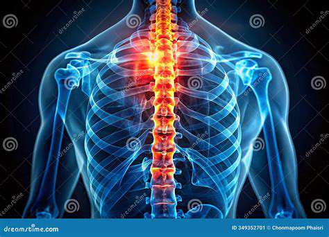
X-rays are a form of electromagnetic radiation with high energy and short wavelengths. They are generated when high-speed electrons are abruptly decelerated, typically within a vacuum tube. This process creates electromagnetic waves capable of penetrating soft tissues but being absorbed by denser materials like bones.
Understanding the fundamental physics behind X-rays is crucial for comprehending how they interact with different tissues. By varying the intensity and duration of exposure, radiologists can create images with varying degrees of contrast, highlighting differences in tissue density and identifying abnormalities.
Applications in Medical Diagnosis
X-ray imaging plays a vital role in diagnosing a wide range of medical conditions. From fractures and dislocations to pneumonia and tumors, X-rays can provide crucial insights into the patient's health status. The ability to visualize the skeletal system is particularly valuable, allowing for the detection of bone abnormalities, such as osteoporosis or bone cancer. This information assists medical professionals in formulating accurate diagnoses and developing effective treatment plans.
Furthermore, X-rays are essential in identifying foreign objects, such as swallowed items or metallic fragments. They are also used to monitor the progress of certain medical treatments, such as fractures healing or the effectiveness of lung treatments.
Safety Precautions and Considerations
While X-rays offer invaluable diagnostic capabilities, it's important to be mindful of the potential risks associated with radiation exposure. Minimizing radiation exposure is a key concern in modern radiology, and protocols are carefully designed to limit the amount of radiation absorbed by patients. Protective shielding, optimized imaging techniques, and careful consideration of the level of exposure are employed to ensure patient safety.
Radiographers and medical professionals are trained to adhere to strict safety guidelines and utilize appropriate shielding measures to protect themselves and the patients from unnecessary radiation exposure. Continuous advancements in X-ray technology are also dedicated to minimizing radiation dose while maintaining image quality.
Technological Advancements in X-Ray Imaging
Significant advancements have been made in X-ray technology over the years. Digital X-ray systems have replaced traditional film-based systems, allowing for immediate image viewing and manipulation, and reducing the need for repeated exposures. Digital imaging significantly enhances the quality and accessibility of diagnostic information.
Computer-aided detection (CAD) systems are increasingly integrated into X-ray imaging, assisting radiologists in identifying subtle abnormalities that might be missed by the naked eye. These advancements contribute to a more efficient and accurate diagnosis process.
The Future of X-Ray Technology
The future of X-ray technology holds immense promise, with ongoing research focused on minimizing radiation dose while enhancing image quality. Innovations like cone-beam computed tomography (CBCT) and dual-energy X-ray absorptiometry (DEXA) are expanding the scope of X-ray applications, providing even more detailed and specialized information. This constant evolution emphasizes the continued importance of X-rays in modern medical practice.
Further advancements in artificial intelligence (AI) are expected to play an increasingly important role in X-ray image analysis, potentially automating the detection of abnormalities and improving diagnostic accuracy. These advancements promise to revolutionize diagnostic imaging and ultimately enhance patient care.
Ultrasound: Exploring Soft Tissues and Organs
Understanding Ultrasound Technology
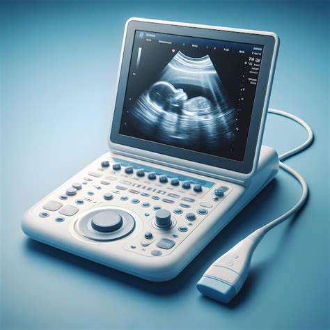
Ultrasound technology, a non-invasive imaging technique, utilizes high-frequency sound waves to create images of the inside of the body. These sound waves bounce off different tissues and organs, returning echoes that a computer interprets to form a visual representation. This technology has revolutionized medical diagnostics, allowing physicians to visualize soft tissues with remarkable clarity and detail. It's a critical tool in various medical specialties, offering valuable insights into the structure and function of internal organs and tissues without the need for harmful radiation.
Applications in Diagnosing Soft Tissue Conditions
Ultrasound plays a crucial role in detecting a wide range of soft tissue conditions. From musculoskeletal injuries like tears and sprains to identifying inflammatory processes and tumors, ultrasound can provide critical information for diagnosis and treatment planning. The ability to image in real-time allows for dynamic observation of the tissues, which is invaluable in assessing the movement and function of organs and structures. This real-time capability is particularly useful in guiding procedures like biopsies and injections.
Imaging Musculoskeletal Structures
Ultrasound is exceptionally well-suited for evaluating musculoskeletal structures. It can visualize tendons, ligaments, muscles, and bursae, enabling the detection of various pathologies such as tears, strains, and bursitis. The precise visualization of these structures allows for accurate diagnosis and helps guide treatment interventions, including physical therapy and surgical procedures. This non-invasive approach is increasingly preferred over other imaging modalities for its accessibility and safety, especially for patients with concerns about radiation exposure.
Evaluating Soft Tissue Tumors and Masses
Ultrasound is a valuable tool for evaluating soft tissue tumors and masses. The ability to differentiate between solid and cystic lesions, combined with the real-time observation of the structures, helps clinicians determine the nature of the abnormality. This information is critical for determining the appropriate course of action, whether it's further investigation, biopsy, or monitoring. This non-invasive imaging technique provides crucial information for making informed treatment decisions.
Guidance for Procedures and Interventions
Ultrasound is frequently used as a real-time guidance tool during various medical procedures and interventions. This includes biopsies, injections, and drainage procedures. The real-time visualization offered by ultrasound ensures precise targeting of the target area, minimizing discomfort and complications. This technology enhances the precision of procedures, improving patient outcomes and reducing the risk of complications.
Safety and Advantages of Ultrasound
Ultrasound is a safe and non-invasive imaging technique. It does not utilize ionizing radiation, making it a preferred option for repeated examinations, especially in pregnant women and young patients. The portability and cost-effectiveness of ultrasound equipment make it accessible in various healthcare settings. This accessibility, coupled with its ability to provide real-time information, makes it a cornerstone of modern medical diagnostics and treatment planning. Furthermore, it offers a high degree of accuracy in depicting soft tissue structures and conditions.

Read more about X Rays and Ultrasounds for Pets: Diagnostic Imaging


Hot Recommendations
- Holistic Pet Health: Integrating Approaches
- The Future of Pet Identification: Biometric Scanners
- Service Dogs for PTSD: A Guide to Support
- The Benefits of Non Anesthetic Professional Teeth Cleaning
- Herbal Supplements for Pet Joint Health
- The Intersection of IoT and Pet Wellness
- Healthy Weight Management for Senior Pets
- The Best Pet Beds for Orthopedic Support and Comfort
- Competitive Dog Sports: Agility, Flyball, Dock Diving
- Luxury Pet Hotels: Pampering Your Beloved Pet